Introduction to Ultrasound Imaging for Lumbopelvic Rehabilitation
An On-Demand Course
About the Course
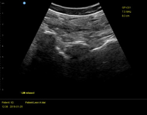
Rehabilitative ultrasound imaging (RUSI) allows for valid, reliable, efficient, and non-invasive measurement of motor control deficits of the deep stabilizing muscles. This continuing education course will provide a basic understanding of how to utilize RUSI for addressing a variety of diagnoses associated with lumbopelvic dysfunction. The material will explore the basic science, clinical evidence, image generation, and sonographic anatomy for the evaluation of tissue morphology and training the muscles of local control.
The course format includes a list of RUSI resources and terminology. Lectures on basic ultrasound science, image generation, and various clinical applications. Each lecture is followed by technique demonstrations that display a split-screen, showing the ultrasound transducer on the body with a simultaneous view of the ultrasound screen.
Instructional Level
Intermediate clinical experience but beginning level for ultrasound imaging. For scope of practice guidelines participants should consult their professional practice act for limitations or training requirements. Content is not intended for use outside the scope of the learner’s license or regulatory agency
Prerequisites
Licensed health care provider with a working knowledge of musculoskeletal anatomy of the lumbopelvic region. Prior experience with or access to rehabilitative ultrasound imaging equipment is NOT required.
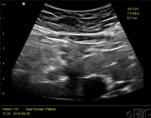
Anterior Abdominal Wall
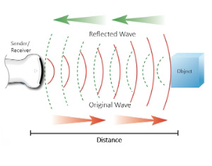
Ultrasound Physics
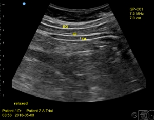
Lateral Abdominal Wall
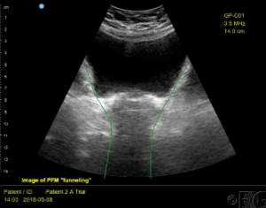
Transabdominal Pelvic Floor
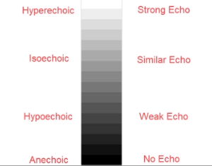
Image Generation
